

We use cookies to ensure our website works properly and to personalise your experience. Cookies policy
Government Institute of Medical Sciences, Greater Noida, India
An anomalous muscle was encountered during the routine dissection of the axillary region of an adult male cadaver by undergraduate students. This slender muscle was present on the right side and was having a small belly with a rounded tendon and oriented transversely in the lower part of axilla. It arose from the upper part of lateral border of latissimus dorsi muscle and extending anteriorly as an arch over the neurovascular bundles present in the axilla. The slender belly of this muscle soon transforms into the rounded tendon and gets inserted into the fascia covering the lower border of the pectoralis major muscle. This variation is described previously as an axillary arch muscle. This anomalous muscle may compress the neurovascular structures producing compression symptoms in the upper limb. So, clinicians and physiotherapists need to know this variation for better diagnosis and treatment.
The axilla is an area of extremely complicated anatomy that is clinically significant. It contains lymph nodes, axillary blood vessels and branches of the brachial plexus [1]. An axillary arch muscle is a muscular or fibro-muscular slip of variable dimensions, extending from the latissimus dorsi muscle to the tendons, muscles, or fasciae surrounding the superior-anterior region of the humerus [2]. Ramsay first characterised the axillary arch muscle in 1795 [3]. The first detailed description of this variation was published in 1846 by Karl Langer Ritter von Edinberg, who noted that the axillary veins, arteries, and nerves that are located beneath this muscle which may become uncomfortable due to the muscle's forceful contractions [4]. The axillary arch muscle is commonly observed in 7–8% of cases [5]. Axillary arch muscle is innervated chiefly by the lateral thoracic nerve. However, its innervation in some cases is from intercostobrachial, internal thoracic, and thoracodorsal nerves. It can be unilateral or bilateral, completely muscular or fibrotic, and have one or more connection points [6]. The axillary arch muscle does not have much functional importance; however, a small study on 22 cases addressed increased strength, motor control of the arm with this variation compared to those without the variation [7]. The importance of this variation is due to its proximity to the important anatomical structures present in the axilla. The palpation of this muscle can be similar to the palpation of enlarged soft tissue or axillary lymph nodes and cause potential problems in armpit surgeries [8]. We are reporting a unilateral Axillary Arch muscle in the right axilla which is arching over the neurovascular structures of this region and it seems to be a rare finding in North Indian population.
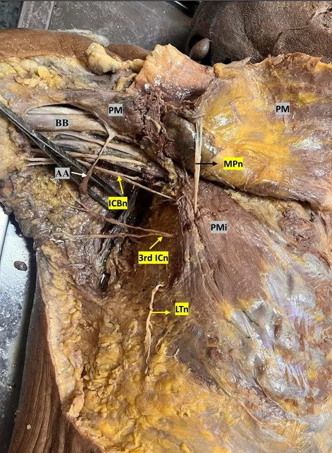
Right Axilla showing axillary arch (AA muscle), BB-Biceps Brachi, PM-Pectoralis major, PMi- Pectoralis Minor, MPn- Median pectoral Nerve, ICBn- Intercostobrachial Nerve, 3rd ICn- Third Intercostal Nerve, LTn-Long Thoracic Nerve.
CASE REPORT:
An anomalous muscle was encountered during the routine dissection of the axillary region of an adult male cadaver by undergraduate students at the Government Institute of Medical Sciences, Greater Noida (UP). This slender muscle was present on the right side had a small belly with a rounded tendon. The axilla, pectoral, scapular regions and the flexor compartment of the arm were exposed by dissecting skin, superficial fascia and deep fascia. The latissimus dorsi, pectoralis major, pectoralis minor, biceps brachii and neuro-vascular structures present in this region were dissected neatly. This anomalous muscular slip arose as fleshy fibers from the lateral border of latissimus dorsi and was oriented transversely in the axilla. This muscular slip was measured approximately about 8 cm in length and 1.5 cm (at the fleshy part) and 0.5 cm (tendinous part) in width. The proximal one third of the muscle was fleshy and the distal two third was tendinous. It traversed anteriorly forming an arch over the neurovascular bundles and gets inserted into the fascia covering the lower border of the pectoralis major muscle. A small twig from Intercostobrachial nerve entered and innervated it from back. The anomalous muscular slip was identified as an axillary arch muscle (Fig: 1). However, the anatomy of these regions on the left side was found normal.
DISCUSSION:
Ramsay (1812) was the first author to observe an oblong muscle that stretched from the pectoralis major to the latissimus dorsi and teres major [3]. Since the first descriptions of the axillary arch muscle were made by Langer (1846), the arch has sometimes been referred to as Langer’s axillary arch [4]. The axillary arch muscle is reported to occur in 7%-8% of dissected cadavers [5]. Usually, the axillary arch muscle shows a bidirectional slip with one origin and one insertion. Few cases have been described where the muscle's connections are more complex, inserting at multiple sites [9]. It was described as a single bi-directional band but can split into a double or multi-band structure extending across the different parts of the axilla [10]. The latissimus dorsi is the consistent source of the Langer's axillary arch. Its insertion occurs on a variety of sites, such as the first rib, teres major, inferior edge of the pectoralis minor, coracobrachialis, long head and short head of the biceps brachii, coracobrachial and axillary fascia. Its dimensions can range from 0.5 to 1.5 cm in width and 7 to 10 cm in length. A branch of the thoraco-dorsal nerve, which also supplied the latissimus dorsi muscle, supplied the muscle12.
We found a unilateral anomalous muscular slip on the right side which arose as separate muscular fibres from the lateral margin of the latissimus dorsi. It was oriented transversely, crossed the neurovascular bundle of the axilla and finally getting inserted into the fascia covering the lower part of the pectoralis major muscle. The proximal one third was muscular and distal two third was tendinous.
Embryologically, the axillary arch muscle takes origin from panniculus carnosus, which is an embryological remnant of extensive sheet present underneath the skin. Panniculus carnosus is well developed in lower mammals in comparison to humans where it is regressed and its functional importance decreases during evolution in favour of wider upper limb mobility and is only evident as muscles like platysma and dartos [11].
It has been described in the previous literature that the presence of the axillary arch muscle is associated with the compression of important neurovascular structures present in the axilla like the axillary vein, musculocutaneous, median, and ulnar nerves. Axillary arch muscle can be palpable during clinical examination and can be confused with enlarged lymph nodes and soft tissue tumors [12]. The axillary arch muscle was also involved in the hyperabduction syndrome [13]. To identify the presence of axillary arch muscle in cases of compression syndrome in the Cervico-axillary region, it is suggested to perform a physical examination, which shows the presence of a mass within the axillary region which is leading to loss of the typical axillary concavity. Though, a physical examination may not always be successful in revealing axillary arch muscle, so it may need magnetic resonance imaging for an accurate diagnosis [14].
CONCLUSION
The axillary arch muscle is less reported in the north Indian population and does not have any significant function but it continues to be an interesting anatomical structure with developmental, evolutionary and clinical significance. It may produce compression symptoms or mimic lymphadenopathy. A thorough understanding of this muscle is essential, particularly when making a clinical diagnosis and carrying out surgical procedures in this area such as breast surgeries. So, it is of immense importance for not only to anatomists but clinicians and physiotherapists also.
REFERENCES

Supreet Kaur*, Unilateral Axillopectoral Muscle – A Case Report And Review Of Its Clinical Significance, Int. J. Sci. R. Tech., 2026, 3 (10), 371-374. https://doi.org/10.5281/zenodo.23163450
 10.5281/zenodo.23163450