

We use cookies to ensure our website works properly and to personalise your experience. Cookies policy
Delight College of Pharmacy, Koregaon Bhima, Pune, Maharashtra, India-412216.
Lung cancer is one of the leading causes of cancer-related mortality worldwide, highlighting the need for effective drug delivery strategies that improve therapeutic outcomes and minimize systemic toxicity. Paclitaxel, a taxane-class anticancer agent, is widely used in selected lung cancer treatment regimens. However, its poor aqueous solubility, formulation-related challenges, systemic adverse effects, and potential for drug resistance limit its therapeutic effectiveness. Nanostructured lipid carriers (NLCs) have emerged as promising second-generation lipid-based drug delivery systems composed of solid lipids and liquid lipids stabilized by surfactants. These carriers offer potential advantages, including improved drug incorporation, enhanced formulation stability, and controlled or sustained drug release. This review focuses on the targeted delivery of paclitaxel using NLCs for lung cancer therapy, emphasizing their composition, structural characteristics, preparation methods, and mechanisms of drug delivery. It also discusses the characterization and quality assessment of paclitaxel-loaded NLCs, including particle size, polydispersity index, zeta potential, encapsulation efficiency, drug-loading capacity, in-vitro drug release, and stability. Furthermore, emerging approaches such as surface-functionalized NLCs, ligand-mediated targeting, pulmonary drug delivery, stimuli-responsive systems, and combination therapy are explored. Despite their potential to improve drug delivery and tumour exposure, challenges related to formulation optimization, long-term stability, large-scale manufacturing, biological variability, and clinical translation remain. Further preclinical and clinical investigations are required to establish the safety, efficacy, and therapeutic advantages of paclitaxel-loaded NLCs in lung cancer management.
1.1. Cancer
Cancer is a disease in which some of the body’s cells grow uncontrollably and spread to other parts of the body. cancer can start almost anywhere in the human body, which is made up of trillions of cells. Normally, human cells grow and multiply (through a process called cell division) to form new cells as the body needs them. When cells grow old or become damaged, they die, and new c ells take their place.
Cancer is a disease of the genome that is both mysterious and terrifying. It is defined by a genomic instability in which many point mutations build up and structural changes take place during the tumor's evolution.1,2 Cancer is a hereditary condition. One significant event in the early phases of tumor formation is the manifestation of oncogenesis.3 Human cells that have, in a sense, gotten away with it and been recruited and partially changed into pathogenic organisms or tumor building blocks are its agents of destruction.1 It is commonly known that inflammatory alterations and the tissue microenvironment directly affect the survival of tumor growth.4 Over the past century, we have made remarkable progress in understanding the biology of cancer. Technological and conceptual advancements in a number of areas, such as massive next-generation sequencing, the inclusion of "omic" sciences, high-resolution microscopy, molecular immunology, flow cytometry, individual cell analysis and sequencing, new cell culture techniques, and the creation of animal models, have been particularly responsible for this progress in recent decades.5 In oncology, personalized (or precision) medicine is based on the concept that tumors differ from one another and that an individual's response to treatment may vary depending on their lifestyle, environment, and genetic profile.6 Chronic inflammatory reactions are essential for the development of inflammation-associated cancers, such as gastric cancer linked to Helicobacter pylori (H. pylori), hepatocellular carcinoma linked to the hepatitis virus, and colon cancer linked to colitis.7 The therapeutic effectiveness of many medications is limited by therapy resistance, and chemoresistance in particular, which leads to relapse and metastasis, according to data from multiple studies.8Cancer has a significant impact on nations of all financial levels. Early detection, better treatment, and a decline in the incidence of known risk factors are all helping to lower the rates of many malignancies in Western nations.9
1.2. Types:
1.2.1 Breast cancer:
Breast cancer is one of the many common malignant tumors that harm women. Numerous internal and external factors can contribute to the development and occurrence of breast cancer.10 Rudolf Virchow postulated a physiological connection between inflammation and ancer in 1863. He postulated that persistent inflammatory areas were the source of cancer. It is now clear that inflammatory cells significantly influence the growth of tumors. Leukocyte infiltration, the expression of cytokines like tumor necrosis factor (TNF) or IL-1, chemokines like CCL2 and CXCL8, active tissue remodeling, and neo-angiogenesis are some of the pro-tumor effects of inflammatory cells. Macrophages linked to tumors have a crucial role in controlling the relationship between inflammation and cancer.11Depending on the related class (i.e., basal-like, Luminal A, Luminal B, HER2-amplified) and the state of molecular markers, distinct breast tumor subtypes receive different treatments. HER2 negative cancers that are positive for the estrogen receptor (ER) and/or progesterone receptor (PR) are usually treated with hormonal therapy as a first line of treatment. These tumors often have a better prognosis than those that are hormone receptor-negative. The American Society for Clinical Oncology does not prescribe a specific chemotherapeutic treatment plan for hormone-positive BC; however, platinum-based medications, taxanes, and anthracyclines are effective alternatives.11 The risk of developing breast cancer increases with age. The majority of female breast cancer cases are identified between the ages of 50 and 69.10
1.2.2. Gastric cancer:
Among lymphomas, sarcomas, gastrointestinal stromal tumors, and neuroendocrine tumors, gastric cancer is the most prevalent stomach cancer. The majority of cases of this diverse disease are gastric adenocarcinomas (GC), which present with a variety of characteristics. The cardia and the non-cardia GC are the two primary GC based on topographic locations.12 Elevated risks of gastric cancer are consistently associated with alcohol consumption and smoking.13
Proximal gastric (cardia) and distal gastric (non-cardia) are the two primary locations of gastric cancer.14 Surgery, chemotherapy, radiation therapy, targeted therapy, and immunotherapy are all part of the treatment options for stomach cancer. Chemotherapy and radiation therapy are frequently utilized as adjuvant or neoadjuvant therapies to improve results, but surgical resection is still the major treatment choice for localized disease.13
1.2.3. Pancreatic cancer:
Usually discovered at an advanced stage, pancreatic cancer is resistant to most treatments, which lowers survival rates. An aging population and rising rates of obesity and pancreatitis, two risk factors for pancreatic cancer, are associated with a steadily rising incidence of the disease.15 The most prevalent chronic liver disease in the world is metabolic dysfunction-associated steatotic liver disease (MASLD), which affects 68% of individuals with type 2 diabetes. However, nothing is known about pancreatic inflammation and fat infiltration in MASLD patients.16 Cigarette smoking, diabetes mellitus, body mass index, alcohol, pancreatitis, allergies, and the role of the microbiome are all known risk factors for pancreatic cancer.17
1.2.4. Oral cancer:
The most common type of cancer in these regions is head and neck cancer, which includes malignancies of the paranasal sinuses, larynx, nasal cavity, pharynx, orbital cavity, and lips. Squamous cell carcinoma (SCC) and cancers of the mouth, head, and neck are closely related. Oral papillomavirus infection (HPV infection) is the term used to describe the human papillomavirus infection that causes oral carcinogenesis.18 From the first rough descriptions of oral neoplasms by a number of significant physicians and surgeons of ancient past civilizations to the current and developing approaches for treating them, oral cancer and its symptoms and signs have been observed and described by medicine since ancient times. All subsites of the oral cavity and oropharynx, as well as malignancies of the lip, are considered oral cancer.19 Patients who receive treatment for early-stage oral cancer have better survival and quality of life rates, as well as a favorable prognosis. However, screening offers a chance for early diagnosis because early-stage malignancies are frequently asymptomatic and mimic benign diseases, which decreases the likelihood that the public will seek care.20 Oral cancer (OC) and other chronic illnesses can significantly affect a person's quality of life. Malignant neoplasms in the oral cavity and oncological treatment frequently lead to reduced oral functions and cosmetic deformity because oral cavity structures execute crucial tasks such communication, mastication, swallowing, and salivation.21Men in their sixth and seventh decades of life are typically affected by oral cancer, and they are more likely to smoke and drink heavily. However, the prevalence of oral cancer in younger patients (less than 45 years old) has increased over the past few decades, rising from 3% to 5% in the 1970s and 1980s to almost 10% currently.22
1.2.5. Prostate cancer:
Beneath the bladder and encircling the urethra is the male reproductive auxiliary organ known as the prostate gland. Contributing vital secretions to semen, which produce ejaculate and preserve sperm viability, is the primary role of the prostate. Most typically in the mid-to-late stage of life, the cells in the prostate gland can develop into tumors.23 Men of all races and ethnicities are susceptible to prostate cancer, and because the disease is often detected late, the mortality rate is higher for those from lower socioeconomic backgrounds. An individual's genetic profile may play a role in prostate cancer, according to mounting evidence.24Hugh Hampton Young published the first significant description of radical prostatectomy using a perineal technique in Annals of Surgery in 1905, marking the beginning of the procedure's usage to treat prostate cancer.25 For many years, research has been conducted on hereditary prostate cancer and a genetic component propensity to prostate cancer. Family inheritance is one of the most significant genetic risk factors for prostate cancer. The relevance of hereditary prostate cancer has been established by both epidemiological and twin investigations.24 With a transperineal approach lowering post-procedural sepsis rates, multimodal early detection using serum prostate-specific antigen (PSA) and multi parametric magnetic resonance imaging (mpMRI) has decreased the requirement for biopsy. More precise staging using PSMA positron emission tomography/computed tomography (PET/CT) prevents unnecessary local interventions, and active surveillance is now a treatment option for localized disease, lowering risks like radiation cystitis and proctitis, erectile dysfunction, and incontinence. When choosing individualized treatment, the patient's characteristics-such as their lifestyle, comorbid illnesses, and symptom profile are the main consideration.26
1.2.6. Gallbladder cancer:
A global issue, gall bladder cancer (GBC) is more common in regions of the world where cholelithiasis is prevalent. The mortality rate for GBC is considerable because it is typically discovered at an advanced stage.27 Along with intrahepaticcholangiocarcinoma (iCCA), extrahepaticcholangiocarcinoma (eCCA), perihilarcholangiocarcinoma (pCCA or Klatskin tumor), and distal cholangiocarcinoma (dCCA), gallbladder cancer (GBC) is classified as a subgroup of biliary tract cancer (BTC).28 A primary care physician in the present day will inevitably meet a variety of disorders that can affect proper gallbladder functioning.29 People all throughout the world, especially those who are at high risk of GBC, now urgently need access to reliable and helpful cancer prevention information in the current environment of varied and frequently contradicting health information. Public health campaigns that educate the public about the risk factors for GBC, including obesity, gallstones, chronic gallbladder inflammation, and factors that contribute to higher mortality rates, such as eating red meat, can help lower the incidence of GBC and improve the prognosis for those who have it.30
1.2.7. Colorectal cancer:
Colorectal cancer (CRC), which includes colon and/or rectum cancer, is a serious health issue as the third most common diagnosed and second most deadly cancer worldwide.31Colon cancer is one of the most deadly cancers, spreading to the liver, lungs, ovaries, and other parts of the gastrointestinal system.32 Due to the anatomical difficulties and the possibility of local recurrence, a more complicated algorithm is required to treat rectal cancer than colon cancer.33 Men have a 25% greater incidence and mortality rate than women, and their 5-and 10-year survival rates are 65% and 58%, respectively. Asian Americans and Pacific Islanders have the lowest incidence and mortality rates of colorectal cancer (CRC), while non-Hispanic Blacks have the highest rates.34 Nowadays, the most widely used CRC serum biomarker is carcinoembryonic antigen (CEA. Both the growing fetus and several cancer cell types produce the protein CEA. CEA levels can be increased in patients with colorectal cancer (CRC), but they are normally low in healthy individuals. Since CEA can also be high in people with other conditions, such as colon inflammation, it is not a precise biomarker for colorectal cancer. Nonetheless, it is a helpful biomarker for tracking the disease's course and recurrence in CRC patients.35
1.2.8. Thyroid Cancer:
Thyroid cancer is the most common endocrine cancer. Thyroid cancer is originating from follicular epithelial cells or parafollicular cells. Follicular cells derived thyroid cancers are classified into 4 histological types: papillary thyroid cancer (PTC80-85%), follicular thyroid cancer (FTC10–15%), poorly differentiated thyroid cancer (PDTC,<2%), and anaplastic thyroid cancer (ATC,<2%).36 Over the pasts several decades, the approach to the diagnosis and management of patients with follicular cell derived thyroid cancer has evolved based on improved classification of patients better matching clinical outcomes, as well as advances in imaging, laboratory, molecular technologies, and knowledge. While thyroid surgery, radioactive iodine therapy, and TSH suppression remain the main stays of treatment, this expansion of knowledge has enabled escalation of therapy for individuals diagnosed with low risk well differentiated thyroid cancer; better definition of treatment choices for patients with more aggressive disease; and improvedability to optimize treatments for patients with persistent and/ or progressive disease.37 There are several known modifiable risk factors for thyroid cancer, however it's not apparent which of these factors is responsible for the rise in thyroid cancer incidence. Patient age, sex, radiation exposure, nutritional status, environmental contaminants, age/ethnicity, and family history comprise the majority of known or suspected risk variables.38
1.2.9. Lung Cancer
Lung cancer is a disease caused by uncontrolled cell division in lungs, usually in the air ways (bronchi or bronchioles) or small air sacs (alveoli). Cells divide make more copies of themselves as a part of their normal function. But sometimes they get changes (mutation) that cause them to keep making more of themselves when they shouldn’t damage cells dividing uncontrollably create masses, or tumours, of tissue. That eventually keeps your organs from working properly39
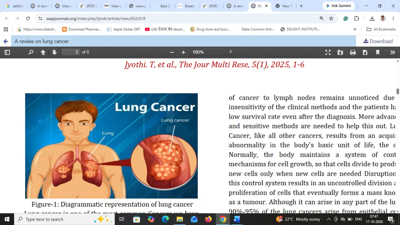
Figure 1: Diagrammatic Representation of Lung Cancer
Lung cancer is one of the most common Cancers we have, and many People die of this disease every year39.Lung cancer is the most important reason of cancer related deaths worldwide, as it is often asymptomatic at the onset of disease and generally discovered at advanced stages. LC is mainly divided into two large groups according to histological subtype as follows; small-cell lung carcinoma and non-small cell lung carcinoma (NSCLC). NSCLC comprises of 85% of all LC cases. Histological sub types of NSCLC include squamous cell carcinoma, large cell carcinoma, and adenocarcinoma. Among these subtypes, adenocarcinoma is the predominant subtype. LC screening via Low-dose Computed Tomography (LDCT) has been found to effective in decreasing mortality, hence the application of this practice is gradually increasing. One of the most important reasons of poor survival rate for LC is due in large part to the delayed diagnosis and advanced stage at the time of diagnosis. Providing an effective early diagnosis, itemized pathogenesis, and appropriate treatment and drug selection at the appropriate stage will be helpful in the optimal treatment of LC. Therefore, diagnosis of LC cancer at the earliest stage is highly vital, particularly in high-risk groups where the identification of new biomarkers is urgently needed40.Cancer is not just one disease, but a generic term used toencompass a group of more than two hundred diseases sharing common characteristics. Cancers (carcinomas) are characterized by their unregulated growth and spread of cells to other parts of the body Lung cancer is one of the most common cancers we have anda large number of people die of this disease every year. The disease is often discovered in a late stage, but also in earlier stages lung cancer patients have worse outcome than patients with other cancers. Even without spreading to other organs, during stage I, the survival rate of lung cancer is under 70%. In comparison, for example, breast cancer there is 95% survival in stage41.
Lung cancers arise through a multistep process involving many genetic and epigenetic changes that includes damage of many key cell-cycle genes41.
1.Early detection:
Promote screening programs for high-risk populations to identifying lung cancer at an early age.
2.Treatment advancement:
Support research into new therapies, including targeted treatments and immunotherapy.
3.Smoking cessation: Implement programs to reduce smoking rates, as tobacco use is the leading cause of lung cancer.
4. Effective Treatment: Personalized treatment: Tailoring treatments (surgery, chemotherapy, radiation, immunotherapy) based on the genetic profile of the tumor.
5. Improving Prognosis and Survival Rates: Extended survival rates: Research into treatments and therapies that can increase the life expectancy of patients.
Histopathological classification of lung cancers is based on cellular and molecular subtypes, which is an essential part of diagnosing and managing lung cancers. The 2021 World Health Organization (WHO) classification system of lung tumors divides the lung cancers as follows:42-43
According to the WHO, identifying histologic features, measuring the depth of invasion, and mode of spread is of prognostic value. For example, they state that tumor spread through air spaces is associated with a higher recurrence rate after limited resections and should be reported on pathological evaluation. Also, the previously described clear cell, rhabdoid, and signet ring subtypes have been discontinued by the most recent WHO classification as they appear to be cytologic features that can occur in anyadenocarcinomas. In the 2015 WHO classification system, poorly differentiated carcinomas were reclassified as squamous cell carcinomas if they had p40 expression; as adenocarcinomas with solid subtype if they had thyroid transcription factor 1 expression;and neuroendocrine carcinomas if they had chromogranin and synaptophysin positivity44.
There are two types of lung cancer
1.Non-small cell lung cancer
2.Small cell lung cancer
1. Non-Small Cell Lung Cancer
Non-small cell lung cancers account forapproximately 85% of all lung cancers and is the most common form of lung cancer in both men and women as well as smoke and nonsmokers.
NACLC is further types into three subtypes45
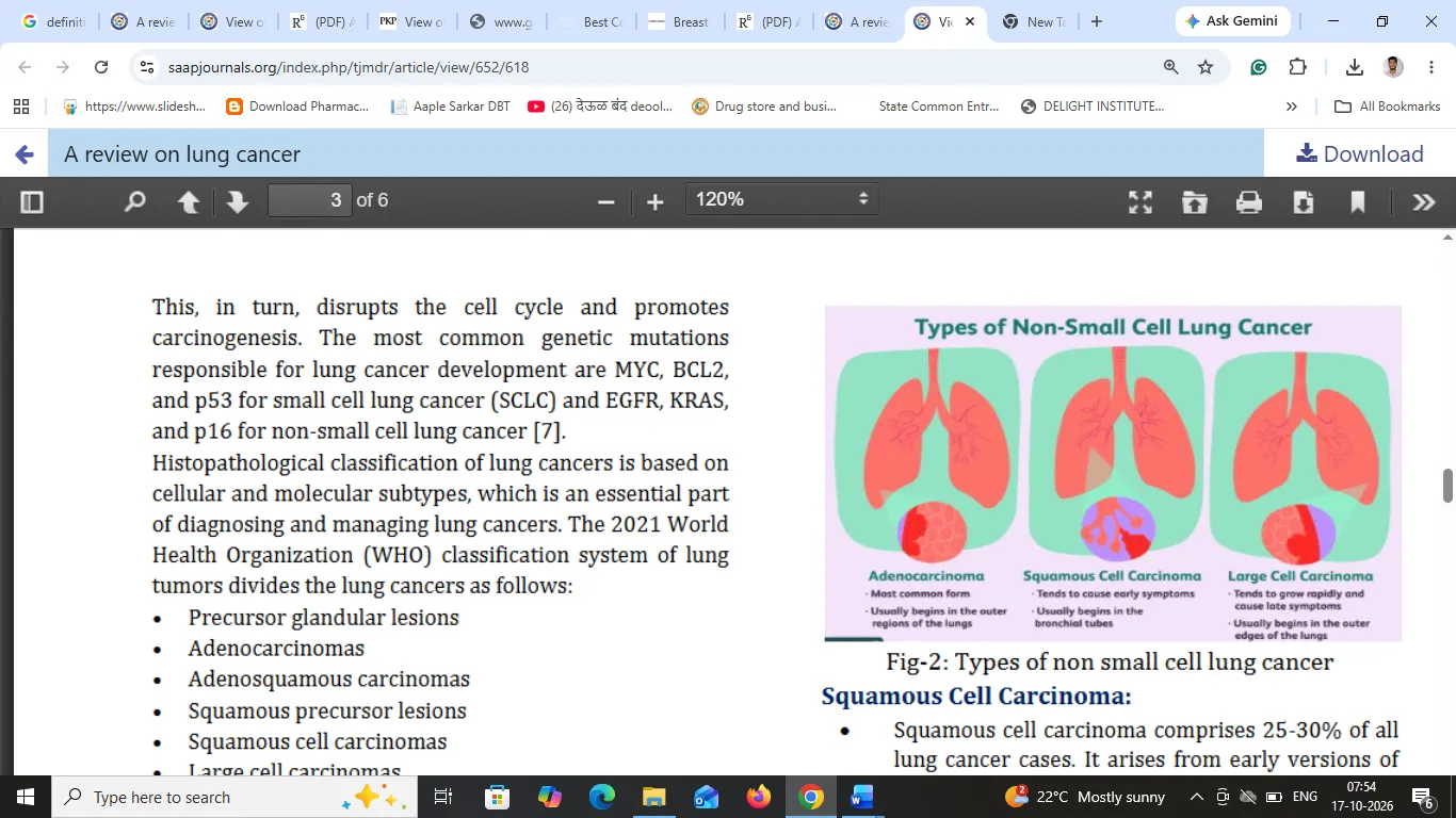
Figure 2: Types of Non-Small Cell Lung Cancer
Types of non-small cell lung cancer
a) Squamous Cell Carcinoma:
Squamous cell carcinoma comprises 25-30% of all lung cancer cases. It arises from early versions of squamous cells in the airway epithelial cells in the bronchial tubes in the center of the lungs. This subtype of NSCLC is strongly correlated with cigarette smoking. This article discusses squamous cell carcinoma in the lungs and its symptoms and causes. It also presents information about diagnosis and treatment45.
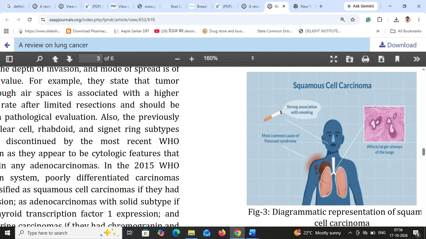
Figure 3: Diagrammatic Representation of Squamous Cell Carcinoma
b) Adenocarcinoma
Stages of Cancer
In ‘Stage zero’ non-small cell lung cancer, abnormal cells are found only in the outer lining of the cells of the lungs. The cancer has not spread to the inner tissues of the lungs, the lymph nodes, or anywhere else in the body. stage zero non-small cell lung cancer is typically treated with surgery
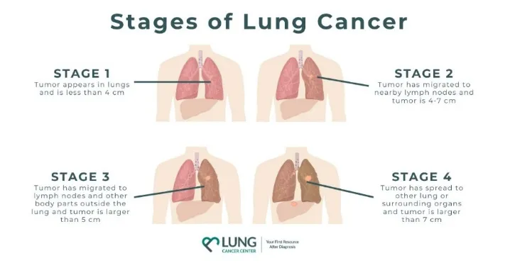
Figure 4: Stages of Cancer
Causes
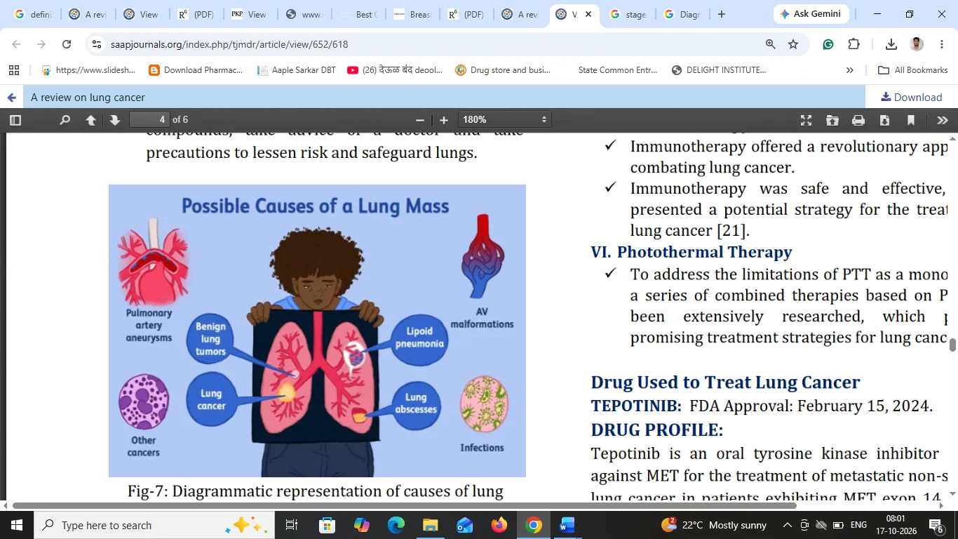
Figure 5: Diagrammatic Representation of Causes of Lung Cancer
Treatment
a) Chemotherapy:
b) Surgery:
c) Adjuvant:
d) Radio Therapy
e) Immuno Therapy
f) Photothermal Therapy
To address the limitations of PTT as a monotherapy, a series of combined therapies based on PTT have been extensively researched, which provided promising treatment strategies for lung cancer47.
2. Nanostructured Lipid Carriers (NLCs)
2.1. Definition
Nanostructured Lipid Carriers (NLCs) are second-generation lipid-based drug delivery systems consisting of a mixture of solid lipids and liquid lipids, stabilized by surfactants. They are generally prepared in the nanometer size range of approximately 50–1000 nm and are designed to improve drug solubility, stability, bioavailability, and controlled drug release.48
2.2. Structure of NLCs
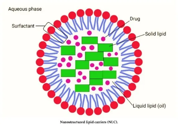
Figure 6: Structure of NLCs
The main structural components are:
2.3. Composition of NLCs
Table I: Composition of NLCs
| Component | Examples | Function |
|---|---|---|
| Solid lipid | Glyceryl monostearate, stearic acid, Compritol 888 ATO | Forms the solid matrix |
| Liquid lipid | Oleic acid, Capryol 90, medium-chain triglycerides | Improves drug incorporation |
| Surfactant | Tween 80, Poloxamer 188, lecithin | Stabilizes nanoparticles |
| Drug | Paclitaxel or other suitable drugs | Provides therapeutic activity |
| Aqueous phase | Purified water or buffer | Disperses the nanoparticles |
2.4. Types of NLCs:
Type I: Imperfect crystal model
The mixture of solid and liquid lipids creates structural imperfections that provide additional space for drug incorporation.
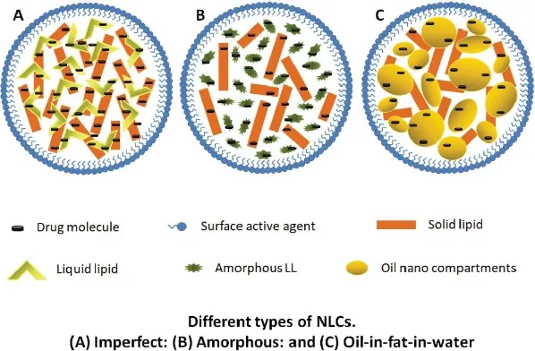
Figure 7: Imperfect Crystal Model
Type II: Multiple type
Small oil-rich domains are dispersed within the solid lipid matrix, helping accommodate drugs with high affinity for the liquid lipid.
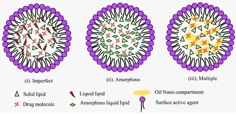
Figure 8: Multiple Type
Type III: Amorphous type
The lipid matrix is designed to reduce crystallization, which can help minimize drug expulsion during storage.
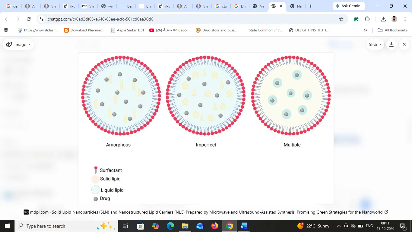
Figure 9: Amorphous Type
2.5. Methods of preparation of NLCs
2.5.1. High-pressure homogenization
Lipids are melted, the drug is incorporated, and a hot aqueous surfactant phase is added. The mixture is homogenized under high pressure and cooled to form nanoparticles.49
2.5.2. Ultrasonication method
The lipid and aqueous phases are mixed and treated with ultrasonic energy to reduce particle size.
2.5.3. Solvent emulsification–evaporation
Lipids and drug are dissolved in a suitable organic solvent, emulsified in an aqueous phase, and the solvent is evaporated to produce nanoparticles.
2.5.4. Solvent injection method
A lipid solution in a water-miscible solvent is injected into an aqueous surfactant solution, leading to lipid precipitation and nanoparticle formation. 48
2.6. Evaluation and characterization of NLCs
Table II: Evaluation and Characterization of NLCs
| Parameter | Purpose |
|---|---|
| Particle size and PDI | Determines particle size distribution and uniformity |
| Zeta potential | Assesses surface charge and colloidal stability |
| Entrapment efficiency (%) | Measures the proportion of drug incorporated into the carriers |
| Drug-loading capacity (%) | Measures the drug content relative to the lipid carrier |
| FTIR | Examines possible chemical interactions |
| DSC and XRD | Evaluate lipid crystallinity and solid-state properties |
| In-vitro drug release | Determines the drug-release profile |
| TEM or SEM | Examines particle morphology |
| Stability studies | Evaluates changes during storage |
2.7. Advantages of NLCs
2.8. Disadvantages of Nanostructured Lipid Carriers (NLCs)
3.Paclitaxel: Complete Drug Profile
3.1. Origin and definition
Paclitaxel is a naturally derived taxane-class anticancer drug originally isolated from the bark of the Pacific yew tree, Taxus brevifolia. It was first identified during the National Cancer Institute's plant-screening programme in the 1960s.
Paclitaxel is a microtubule-stabilizing antineoplastic agent used to treat several cancers, including ovarian cancer, breast cancer and non-small-cell lung cancer (NSCLC).49
3.2. Chemical structure
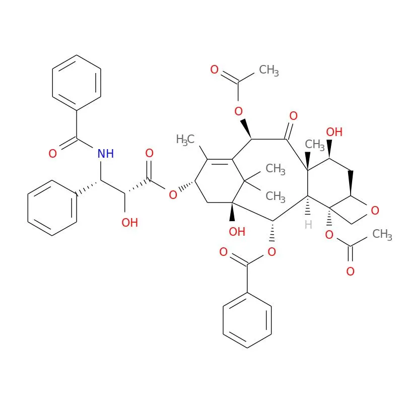
Figure 10: Chemical Structure
Paclitaxel has a complex taxane-ring structure containing an oxetane ring, several hydroxyl groups, ester linkages and a characteristic N-benzoyl phenylisoserine side chain. These structural features are important for its interaction with microtubules.
3.3. IUPAC name (IUPAC chemical nomenclature)
The IUPAC name of paclitaxel is:
[(1S,2S,3R,4S,7R,9S,10S,12R,15S)-4,12-diacetyloxy-15-[(2R,3S)-3-benzamido-2-hydroxy-3-phenylpropanoyl]oxy-1,9-dihydroxy-10,14,17,17-tetramethyl-11-oxo-6-oxatetracyclo[11.3.1.0³,¹â°.0â´,â·]heptadec-13-en-2-yl] benzoate.
3.4. Molecular formula and properties
Table III: Molecular Formula and Properties
| Property | Description |
|---|---|
| Generic name | Paclitaxel |
| Drug class | Taxane anticancer agent |
| Molecular formula | C₄₇Hâ‚…â‚NOâ‚â‚„ |
| Molecular weight | 853.91 g/mol |
| CAS number | 33069-62-4 |
| Physical appearance | White to off-white crystalline powder |
| Solubility | Practically insoluble in water; highly lipophilic |
| Pharmacological action | Microtubule stabilizer |
| Therapeutic class | Antineoplastic agent |
3.5. Mechanism of action (MOA)
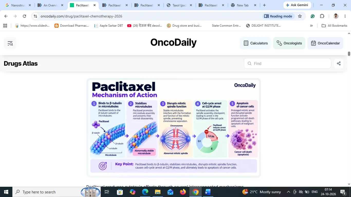
Figure 11: Mechanism of Action (MOA)
Paclitaxel stabilizes microtubules by preventing their depolymerization. This disrupts the dynamic reorganization of microtubules required for mitosis and other cellular functions. The resulting mitotic arrest can lead to apoptosis (programmed cell death) in susceptible cancer cells.50
3.6. Route of administration
The principal conventional route of administration for paclitaxel is intravenous (IV) infusion.
3.7. Conventional formulation of paclitaxel
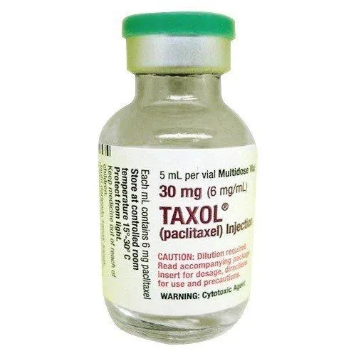
Figure 12: Formulation of Paclitaxel
The conventional formulation of paclitaxel is a sterile concentrate for intravenous infusion.
Table IV: Conventional Formulation of Paclitaxel
| Component | Function |
|---|---|
| Paclitaxel | Active pharmaceutical ingredient (API) |
| Polyoxyethylated castor oil (Cremophor EL) | Solubilizing vehicle for poorly water-soluble paclitaxel |
| Dehydrated ethanol | Cosolvent that helps dissolve paclitaxel |
| Diluent for infusion | Used to prepare the final infusion according to product instructions |
3.8. Limitations of conventional paclitaxel formulation
1. Poor aqueous solubility: Paclitaxel is highly lipophilic and practically insoluble in water.
2. Vehicle-related toxicity: Cremophor EL can contribute to hypersensitivity reactions and other infusion-related adverse effects.
3. Premedication requirements: Conventional formulations generally require premedication according to prescribing information to reduce hypersensitivity risk.
4. Systemic toxicity: Adverse effects can include peripheral neuropathy, myelosuppression and other dose-limiting toxicities.
5. Formulation limitations: The conventional solvent-based formulation may complicate delivery to specific tissues and does not inherently provide targeted drug delivery.51
4.Targeted Delivery of Paclitaxel Using Nanostructured Lipid Carriers (NLCS) For Lung Cancer Therapy
4.1. Introduction
Lung cancer represents the abnormal growth of cells in lung; it is a deadliest cancer globally and remains one of the leading causes of cancer-related mortality worldwide, with adenocarcinoma representing the most prevalent histological subtype. There is various cause of lung cancer but foremost cause is carcinogenic compounds Benzo [α]pyrene, found in tobacco smoking and industrial emission. Benzo [a]pyrene (B [a]P) is a prototypical polycyclic aromatic hydrocarbon (PAH) widely detected in environmental matrices including air, surface water, soil, and sediments. It is also present in cigarette smoke and certain food products, particularly those subjected to smoking or grilling, making human exposure frequent and unavoidable. Increasing evidence highlights the toxicological risks associated with B [a]P. Following exposure, this xenobiotic undergoes metabolic activation by cytochrome P450 enzymes to form the carcinogenic metabolite 7β,8α-dihydroxy-9α,10α-epoxy-7,8,9,10- tetrahydrobenzo [a]pyrene (BPDE). BPDE interacts with DNA to enerate adducts, thereby inducing mutations and promoting malignant transformation. It induces mutation in normal cell and prone to activate abnormal cell as well as it has DNA adduct, so it represents as cancer contributors. It triggers the genetic mutation and surpasses the apoptosis process to contribute lung carcinogenesis and prone to uncontrolled cell proliferation within the lungs. 52
4.2. NLC-Based Paclitaxel Delivery
4.2.1 Overview of Nanostructured Lipid Carriers
NLCs are colloidal drug delivery systems designed to improve the incorporation and delivery of therapeutic agents. Their lipid matrix contains solid and liquid lipids, while surfactants help maintain dispersion stability. Compared with conventional solid lipid nanoparticles, the mixed lipid matrix can provide additional space for drug incorporation and may reduce drug expulsion during storage.
4.2.2 Formulation of Paclitaxel-Loaded NLCs
Paclitaxel-loaded NLCs can be prepared by selecting suitable solid lipids, liquid lipids, surfactants, and processing conditions. Common preparation methods include high-pressure homogenization, ultrasonication, solvent emulsification–evaporation, and solvent injection. Formulation optimization is necessary to achieve appropriate particle size, drug-loading capacity, encapsulation efficiency, stability, and drug-release characteristics.
4.2.3 Advantages of NLC-Based Paclitaxel Delivery
NLCs may improve paclitaxel incorporation, protect the drug from degradation, and provide controlled or sustained release. Depending on their composition and route of administration, they may also be engineered to improve tumour exposure and reduce exposure of healthy tissues. However, these benefits require experimental validation and cannot be assumed for every formulation.53
4.3. Role of NLCs in Paclitaxel Delivery
NLCs may serve as carriers for paclitaxel by incorporating the drug within a lipid matrix and facilitating its transport through biological environments. Their potential roles include:
The performance of NLCs depends on their physicochemical properties, the administration route, the biological environment, and the characteristics of the tumour.53
4.4. Mechanism of NLC-Mediated Drug Delivery to Tumours
NLC-mediated drug delivery refers to the processes through which nanostructured lipid carriers transport an encapsulated drug through the body and facilitate its release at or near the intended site of action.
4.4.1 Administration and Distribution
Following administration, paclitaxel-loaded NLCs enter the relevant biological environment. For intravenous administration, their distribution depends on particle characteristics, protein adsorption, circulation time, and interactions with the mononuclear phagocyte system.
4.4.2 Tumour Accumulation
Some intravenously administered nanoparticles may accumulate in tumours through altered tumour vasculature and impaired lymphatic drainage, commonly described as the enhanced permeability and retention (EPR) effect. This phenomenon varies considerably among tumour types and patients, and its clinical contribution should not be overstated.
4.4.3 Cellular Uptake
NLCs that reach tumour tissue may interact with cancer cells and be internalized through endocytosis or related cellular uptake pathways. Surface modification with suitable targeting ligands may enhance interactions with selected cells when the target is appropriately expressed.
4.4.4 Intracellular Drug Release
After cellular uptake, paclitaxel may be released from the lipid matrix through diffusion, lipid-matrix reorganization, and degradation or digestion of lipid components. Released paclitaxel can interact with β-tubulin, stabilize microtubules, disrupt mitotic spindle function, and inhibit cancer-cell proliferation.
4.4.5 Potential Therapeutic Benefits
The intended outcomes include improved drug delivery to tumour tissue, modified release, and potentially reduced systemic exposure. These effects depend on the formulation and require evaluation using appropriate in-vitro, in-vivo, and clinical studies.
5. Characterization and Quality Assessment of Paclitaxel-Loaded NLCs
Comprehensive characterization is essential to establish the quality, reproducibility, stability, and performance of paclitaxel-loaded NLC formulations.
5.1 Particle Size, Polydispersity Index, and Zeta Potential
Dynamic light scattering is commonly used to determine particle size and polydispersity index (PDI). Zeta potential provides information about surface charge and may help assess colloidal stability.
5.2 Encapsulation Efficiency and Drug-Loading Capacity
Encapsulation efficiency determines the proportion of the initially added drug that is entrapped within or associated with the carrier. Drug-loading capacity measures the amount of drug relative to the carrier or total formulation, depending on the stated calculation method.
5.3 Morphological Evaluation
Transmission electron microscopy (TEM), scanning electron microscopy (SEM), or atomic force microscopy (AFM) may be used to evaluate particle morphology and surface characteristics.
5.4 Solid-State Characterization
Differential scanning calorimetry (DSC), X-ray diffraction (XRD), and Fourier-transform infrared spectroscopy (FTIR) may be employed to investigate lipid crystallinity, the physical state of paclitaxel, and potential interactions between formulation components.
5.5 In-Vitro Drug Release
In-vitro release studies evaluate the rate and extent of paclitaxel release under defined experimental conditions. Suitable validated methods and sink conditions are important for meaningful comparison between formulations.
5.6 Stability Studies
Stability studies assess changes in particle size, PDI, drug content, encapsulation efficiency, appearance, and drug-release characteristics during storage. These studies help establish appropriate storage conditions and a potential shelf life.
5.7 Biological Evaluation
Cellular uptake, cytotoxicity, apoptosis, haemocompatibility where relevant, and other safety endpoints may be evaluated using appropriate models. In-vivo studies may assess pharmacokinetics, biodistribution, tumour response, pulmonary tolerability, and systemic toxicity, depending on the intended route.48
6. Recent Advances and Future Perspectives
Recent research in lipid-based nanocarriers has explored surface-functionalized NLCs, ligand-mediated targeting, inhalable nanocarrier systems, stimuli-responsive formulations, and combination drug delivery. These approaches may offer opportunities to improve the delivery of paclitaxel to lung tumours.
6.1 Surface-Modified and Ligand-Targeted NLCs
Functionalization with selected ligands may improve recognition and uptake by cells expressing the corresponding targets. The effectiveness of such systems depends on ligand density, target expression, particle stability, and biological accessibility.
6.2 Pulmonary Drug Delivery
Inhalable formulations are being investigated to deliver anticancer agents directly to the respiratory tract. For paclitaxel-loaded NLCs, formulation design must address aerosol performance, aerodynamic particle size, lung deposition, mucociliary clearance, pulmonary toxicity, and stability during aerosolization.
6.3 Stimuli-Responsive Delivery
NLC formulations may be designed to respond to specific environmental conditions, such as pH or redox differences, to modify drug release. Their practical value depends on the strength, selectivity, and reproducibility of the response under biologically relevant conditions.
6.4 Combination Therapy
NLCs may provide a platform for co-delivery of paclitaxel and other therapeutic agents. Such combinations require careful assessment of drug compatibility, loading, release kinetics, therapeutic benefit, and toxicity.
6.5 Challenges and Future Directions
Future research should prioritize reproducible manufacturing, long-term stability, scalable production, rigorous safety evaluation, and clinically relevant lung cancer models. A key challenge is demonstrating that improved nanocarrier characteristics translate into meaningful therapeutic benefits in patients.
CONCLUSION
Nanostructured lipid carriers represent a promising platform for investigating the delivery of paclitaxel in lung cancer therapy. Their mixed lipid matrix may improve drug incorporation, modify drug release, and support the development of targeted or pulmonary delivery systems. Nevertheless, challenges related to formulation stability, manufacturing scale-up, biological variability, and clinical translation remain. Further systematic preclinical and clinical investigations are required to establish the safety, efficacy, and therapeutic advantages of paclitaxel-loaded NLCs compared with conventional formulations.
8. Acknowledgement
The author gratefully acknowledges the institutional support and academic resources that contributed to the preparation of this review article. The author also acknowledges the contributions of researchers whose published work has advanced the understanding of nanostructured lipid carriers and paclitaxel-based drug delivery systems.
REFERENCES



Ashwarya R. Gavhane, Vishal S. Madankar, Sampat D. Navale, Targeted Drug Delivery of Paclitaxel Using Nanostructured Lipid Carrier for Lung Cancer Therapy, Int. J. Sci. R. Tech., 2026, 3 (11), 442-457. https://doi.org/10.5281/zenodo.24000416
 10.5281/zenodo.24000416